EMG-Atlas is a specialized instructional resource designed for physicians to improve precision and safety during needle electromyography (EMG) for assessing muscle electrical activity.
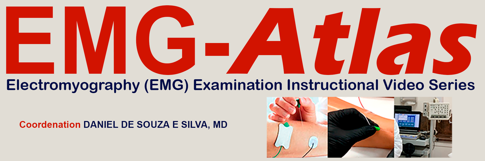
EMG-Atlas is a instructional video series created by Daniel de Souza e Silva, MD and is designed exclusively for physicians and aims to demonstrate the correct technique for needle placement in various muscles during an electromyography (EMG) examination. Each video in the series presents a detailed step-by-step guide, showing the insertion of needles into different muscles of the patient.
EMG-Atlas features 73 muscles across the lower limbs, upper limbs, and trunk;
Real-time synchronization with the examination equipment screen for all muscles included in EMG-Atlas;
High-definition videos showing in detail, with clear instructions, the correct placement of the needle in each muscle examined;
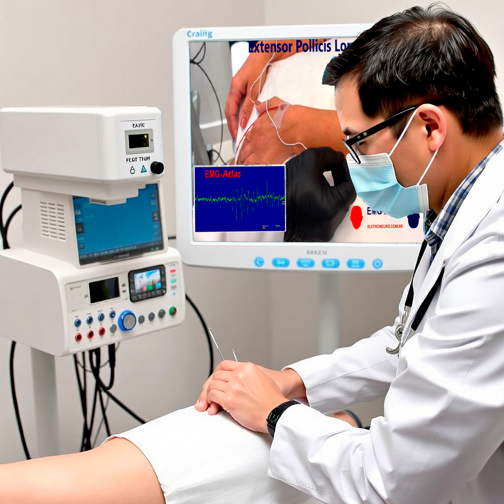
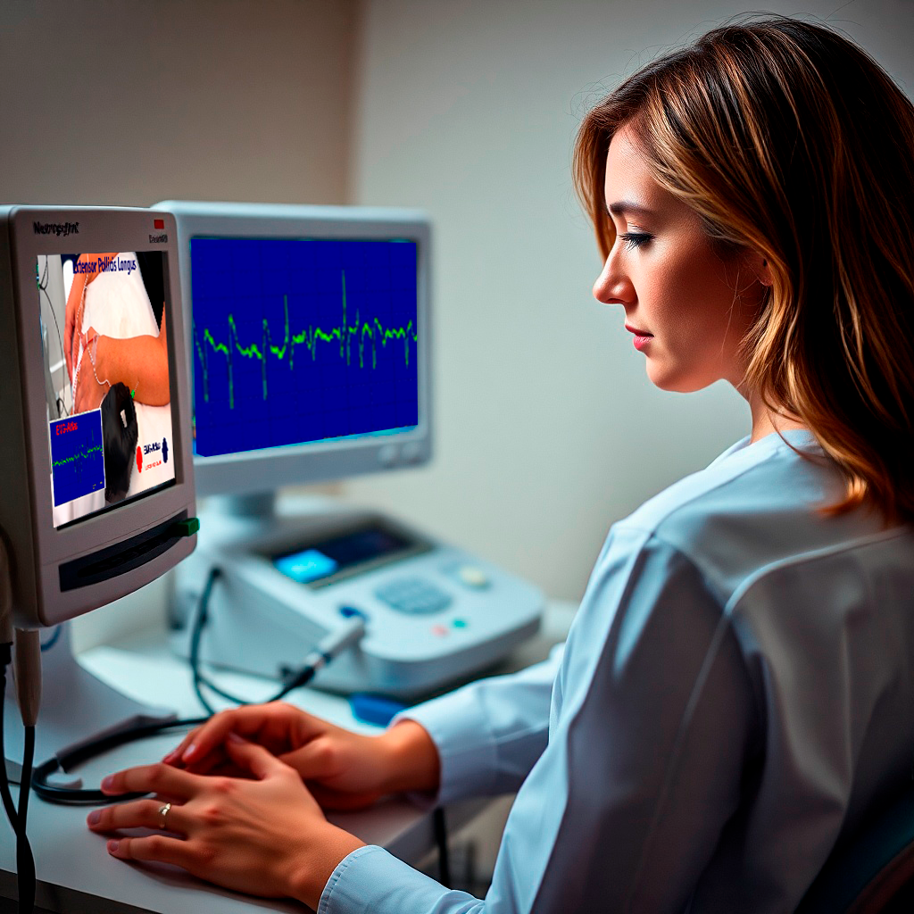
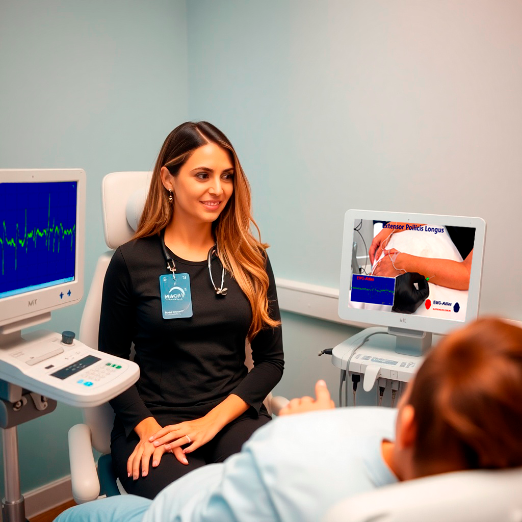
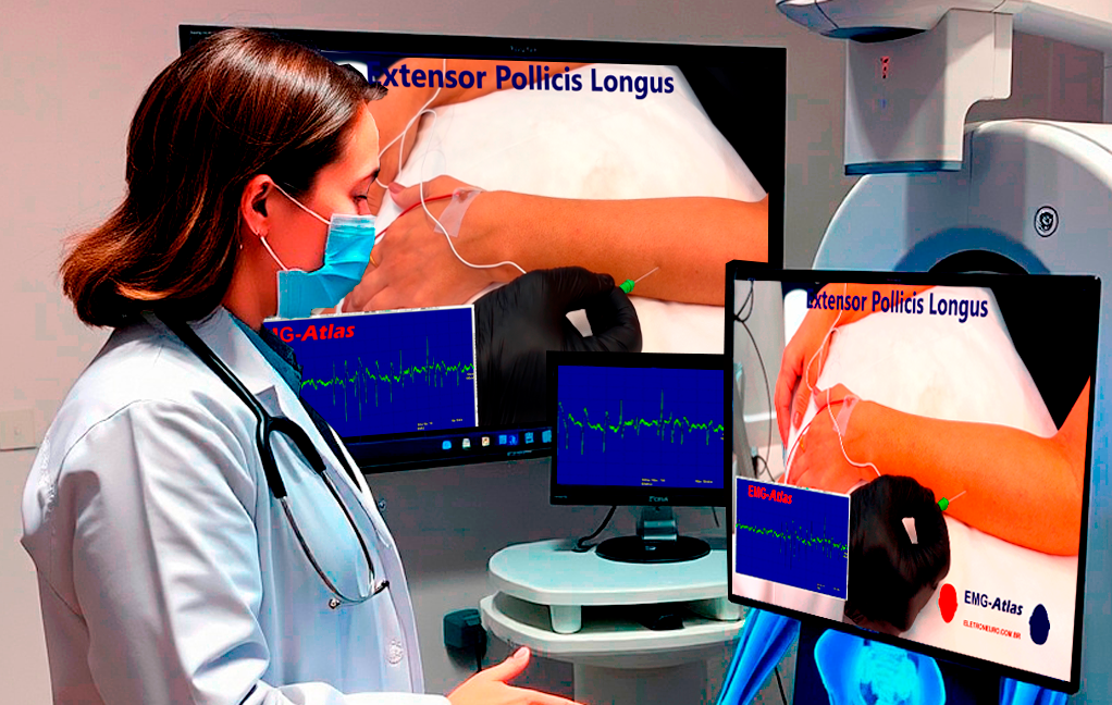
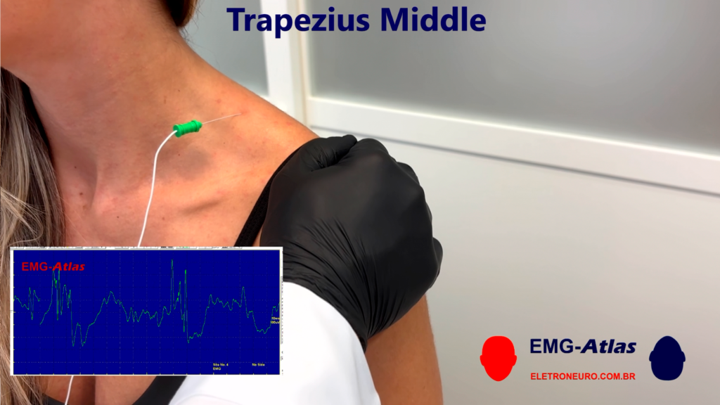
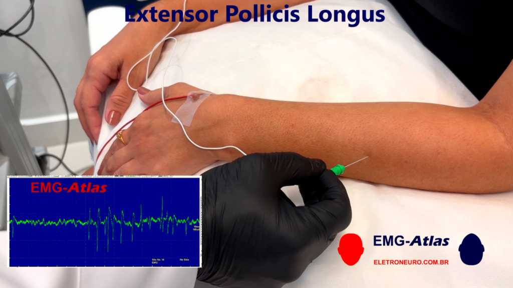
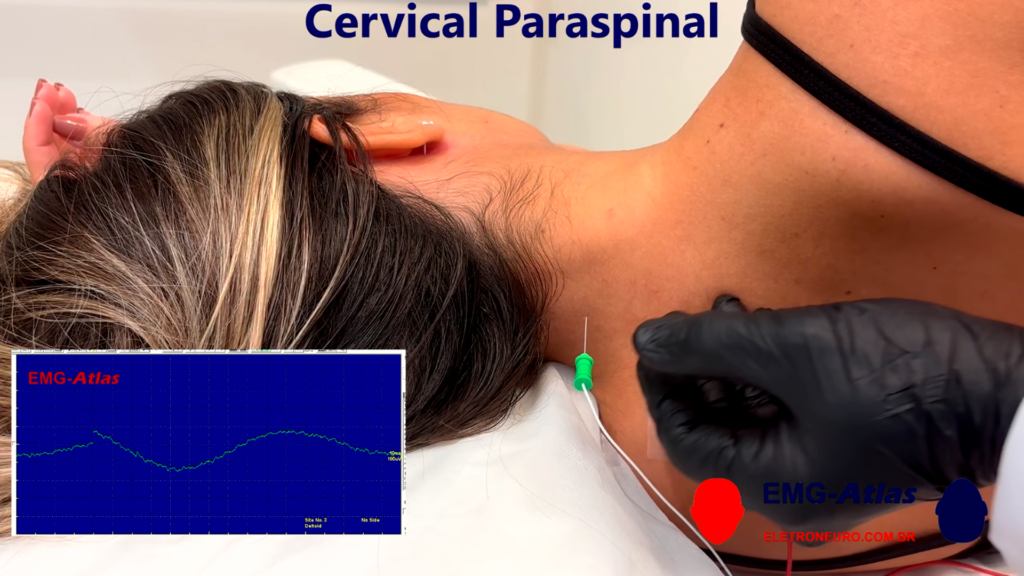
During the videos, in addition to the practical demonstration of needle placement, the electromyography device screen is displayed in real time, synchronized with the patient’s examination. This allows physicians to simultaneously observe the insertion technique and the results obtained on the device.
The videos cover a wide range of muscles, providing specific instructions for each one, and highlight important points such as relevant anatomy, needle insertion depth, and precautions to be taken to ensure the safety and effectiveness of the examination.
This series is a valuable tool for physicians who wish to improve their electromyography skills, offering a practical and detailed overview of the procedure.
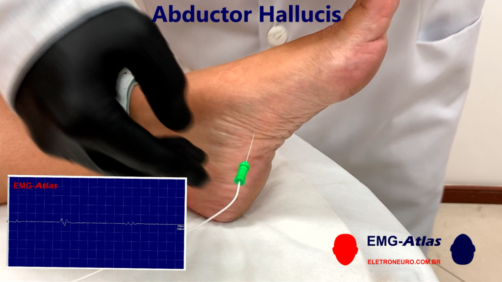
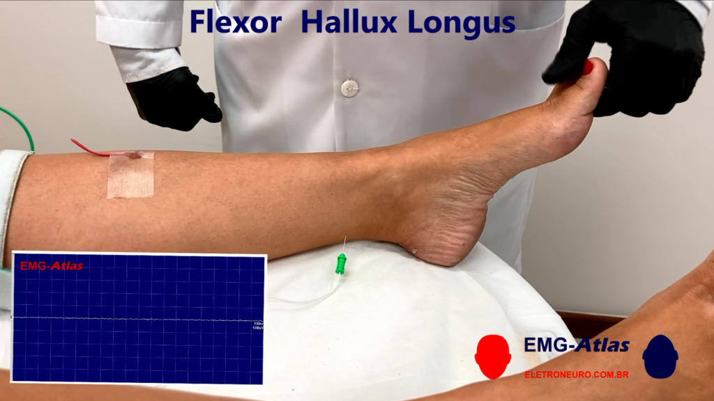
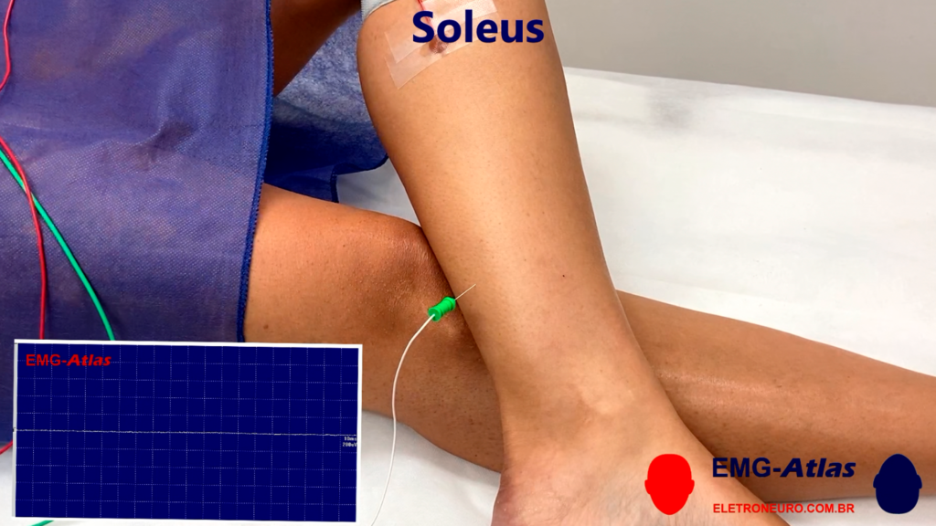
Some EMG-Atlas screenshots:
Mechanisms of Guidance
Step-by-Step Needle Insertion Videos:
EMG-Atlas provides high-definition, real-time video demonstrations showing the insertion of needles into specific muscles.
Each video focuses on a particular muscle and displays the correct trajectory, insertion depth, and anatomical landmarks.
Example muscles covered include the Abductor Pollicis Brevis, Pronator Quadratus, Flexor Digitorum Profundus, Biceps Brachialis, and many others across the upper limb, lower limb, and trunk.
Real-Time Synchronization with EMG Device:
During demonstrations, the EMG device screen is shown synchronized with the needle insertion,
allowing physicians to correlate electrical activity signals with precise needle placement.
Detailed Anatomical Guidance:
EMG-Atlas highlights surface landmarks, bony prominences, and cross-sectional anatomy, essential
for safe and precise needle location.
It emphasizes muscle depth, relative position to tendons and nerves, and variations in muscle
anatomy, aiding in targeting deep or small muscles while avoiding vascular or neural structures.
Activation and Maneuver Instructions:
Physicians are shown active maneuvers for the patient to elicit optimal muscle activation, which
helps confirm correct needle placement.
For instance, the pronation/supination of the forearm or finger flexion may be used to distinguish
amongst muscle slips innervated by different nerves.
Safety and Clinical Considerations:
EMG-Atlas educates on precautions such as avoiding excessive depth, lateral or medial misplacement, and surrounding artery or nerve injury.
Videos highlight patient positioning, insertion angles, and palpation techniques to optimize
comfort and reduce procedural complications.
Comprehensive Muscle Coverage:
The atlas covers 73 muscles in total, with specific guidance for needle insertion points,
direction, and depth.
It bridges theoretical knowledge with practical skills, enabling physicians to refine their
technique through visual and procedural reinforcement.
Specific content and Duration:
Part 1 – Upper Limbs and Trunk includes the following muscles:
- Abductor Digiti Minimi
- Abductor Policis Brevis
- Adductor Pollicis
- Biceps Brachialis
- Brachialis
- Brachioradialis
- Cervical Paraspinal
- Deltoid
- Extensor Carpi Radialis
- Extensor Carpi Ulnaris
- Extensor Digitorum Comunis
- Extensor Indicis Propius
- Extensor Pollicis Brevis
- Extensor Pollicis Longus
- First Dorsal Interosseus
- Flexor Carpi Radialis – Palmaris Longus
- Flexor Carpi Ulnaris
- Flexor Digirotum Profundus
- Flexor Digitorum Sublimis
- Flexor Pollicis Brevis
- Flexor Pollicis Longus
- Infraspinatus
- Latissimus Dorsi
- Opponens Pollicis
- Pectoralis Major
- Pronator Quadratus
- Pronator Teres
- Rhomboideus Major
- Serratus Anterior
- Sternocleidomastoid
- Supinator
- Supraspinatus
- Teres Minor
- Trapezius Middle
- Trapezius Upper
- Triceps Brachialis
TOTAL: (24:31s)
Part 2 (Lower Limbs and Trunk) includes the following muscles:
- Abductor Digiti Quinti
- Abductor Hallucis
- Adductor Brevis
- Aductor Longus
- Adductor Magnus
- Biceps Femoris Long Head
- Biceps Femoris Short Head
- Extensor Digitorum Brevis
- Extensor Digitorum Longus
- Extensor Hallucis Longus
- External Oblique
- Flexor Digitorum Brevis
- Flexor Digitorum Longus
- Flexor Hallucis Brevis
- Flexor Hallucis Longus
- Gastrocnemius Lateral Head
- Gastrocnemius Medial head
- Gluteus Maximus
- Gluteus Medius
- Gluteus Minimus
- Iliopsoas
- Lumbar Paraspinal
- Peroneus Brevis
- Peroneus Longus
- Quadratus Plantae
- Rectus Abdominis
- Rectus Femoris
- Semimembranosus
- Semitendinosus
- Soleus
- Tensa Fascia Lata
- Thoracic Paraspinal
- Tibialis Anterior
- Tibialis Posterior
- Vastus Intermedius
- Vastus Lateralis
- Vastus Medialis
Au fil des semaines, https://bdmbet-frr.com/ confirme ses promesses et fidélise une communauté de joueurs grandissante. Les notifications discrètes alertent sur les événements importants sans détourner l’attention. Le service de caisse propose un suivi en temps réel de chaque opération initiée par le joueur. Le chiffrement SSL 256 bits protège toutes les communications sensibles entre le navigateur et les serveurs. Les variantes Hold’em proposées en live attirent les amateurs de poker traditionnel. Les sessions multi-appareils reprennent leur état exact en passant du desktop au smartphone. Une page d’historique recense les bonus passés, présents et à venir pour davantage de transparence. Les fonctionnalités sociales permettent de partager certains résultats marquants sans contraintes. Les variantes régionales de roulette et de blackjack ouvrent l’horizon vers des styles parfois méconnus en Europe. Le centre d’aide regroupe une foire aux questions copieuse, structurée par thématiques. L’ensemble forme un projet abouti qui semble parti pour durer dans la sphère francophone.
Les joueurs qui s’attardent sur Casino Together y découvrent souvent des recoins méconnus mais bien construits. Les transactions par carte bancaire s’effectuent instantanément avec un crédit immédiat sur le compte du joueur. Les sessions interrompues par un changement de réseau reprennent automatiquement à leur point d’arrêt. Des dossiers thématiques explorent en profondeur certains genres ou certaines mécaniques de jeu. L’inscription s’effectue en quelques étapes simples, sans formulaire à rallonge. Le chiffrement SSL 256 bits protège toutes les communications sensibles entre le navigateur et les serveurs. Une catégorie sans dépôt propose plusieurs titres à essayer librement. Les délais de connexion à une table libre restent généralement très brefs. Une fois découverts, les nombreux atouts de la plateforme rendent un éventuel retour très probable.
Pour qui souhaite varier ses lieux de jeu habituels, https://mrjoker8.com/ constitue une étape qui mérite d’être tentée. La consommation de données est optimisée pour ne pas peser sur les forfaits limités. Les guides débutants accompagnent les nouveaux venus dans la découverte des règles principales. Les frais éventuels sont signalés en amont sans surprise au moment de la validation. Le centre d’aide reste accessible sans nécessiter de connexion préalable. Les amateurs de slots à volatilité maîtrisée trouvent un large éventail de titres correspondant à des budgets modérés. Les délais de connexion à une table libre restent généralement très brefs. Les surprises occasionnelles, sans communication préalable, alimentent la fidélité de la communauté. Le passage par cette adresse offre un complément intéressant à toute routine de jeu déjà établie.
Découvrir casino prince ali sans préjugés permet d’apprécier toute la subtilité du travail réalisé. Les processus de KYC garantissent une vérification approfondie sans intrusivité excessive. Les fiches d’analyse de jeux livrent des informations détaillées sur les mécaniques et les RTP. L’interface privilégie la lisibilité avec une hiérarchie visuelle claire entre menus, contenus et appels à l’action. Le filtre par fournisseur, par thème et par mécanique simplifie la recherche du jeu idéal. L’authentification biométrique simplifie la connexion sur les appareils compatibles. Les protocoles de chiffrement utilisés correspondent aux standards bancaires européens en vigueur. Une page d’historique recense les bonus passés, présents et à venir pour davantage de transparence. Une plateforme à observer dans la durée, qui pourrait bien s’imposer comme une référence durable.
Les sessions répétées chez casino machance renforcent souvent une impression positive plutôt que de l’éroder. Les politiques de confidentialité respectent strictement le RGPD européen ainsi que les standards internationaux. Les conditions de mise sont visibles sur chaque promotion sans nécessiter de recherche complémentaire. La caisse multidevise permet d’opter pour la devise correspondant à ses habitudes. Une assistance téléphonique est ouverte pour les demandes nécessitant un échange plus approfondi. Les statistiques affichées en surimpression aident à suivre les tendances récentes de chaque table. La page d’accueil met en exergue les promotions et nouveautés dans des bandeaux clairement délimités. Les amateurs de jeux Drop & Win bénéficient d’une rotation hebdomadaire des tournois associés. L’authentification biométrique simplifie la connexion sur les appareils compatibles. Une fois découverts, les nombreux atouts de la plateforme rendent un éventuel retour très probable.
Démarrer une session sur https://winnita-fr.com suffit souvent à comprendre pourquoi ce nom revient dans tant de conversations. Les croupiers en direct mémorisent souvent les habitués et personnalisent leurs interventions. Les sauvegardes redondantes protègent les bases de données contre tout incident matériel. Le service de caisse propose un suivi en temps réel de chaque opération initiée par le joueur. Les retours utilisateurs nourrissent les évolutions futures de la plateforme. Les ventes flash ponctuelles permettent d’acquérir des packs de free spins à tarif réduit. La latence du live casino reste contenue sur la version mobile, ce qui surprend agréablement. Les niveaux d’expérience progressifs offrent une approche ludique de la fidélité. Les widgets latéraux affichent les promotions actives sans saturer la lecture des contenus principaux. Une rubrique cartes à gratter offre une parenthèse rapide et accessible entre deux longues sessions. Un projet sérieux qui mérite d’être considéré pour qui cherche un nouveau point de chute en ligne.
Pour beaucoup, ninecasino est devenu une référence à laquelle on revient régulièrement entre deux découvertes. Aucune application native n’est requise : tout se déroule via le navigateur du smartphone. Les taux de redistribution observés sont publiés à intervalles réguliers dans un esprit de transparence. La page d’accueil met en exergue les promotions et nouveautés dans des bandeaux clairement délimités. Les variantes régionales de roulette et de blackjack ouvrent l’horizon vers des styles parfois méconnus en Europe. Les utilisateurs VIP bénéficient d’un manager dédié avec un canal prioritaire. Le système de points fidélité s’accumule au fil des sessions et se convertit selon des barèmes lisibles. Une rubrique communauté centralise actualités, témoignages et conseils tirés des forums. Les sessions en direct fonctionnent en continu, avec une rotation d’équipes qui assure la disponibilité 24/7. Les retraits crypto se finalisent souvent en moins de trente minutes pour les profils déjà vérifiés. Le passage par cette adresse offre un complément intéressant à toute routine de jeu déjà établie.
L’investissement éditorial visible sur Mystake Casino donne à la plateforme une profondeur peu courante. Les statistiques affichées en surimpression aident à suivre les tendances récentes de chaque table. L’application des plafonds journaliers se fait de manière transparente et configurable. Le tableau de bord centralise l’historique, les bonus actifs et les transactions récentes. Les liens de réinitialisation des mots de passe sont à usage unique et à durée limitée. Le mode hors-ligne partiel permet de naviguer dans le compte même en perte temporaire de connexion. Les agents identifient leur fonction et s’expriment avec professionnalisme à chaque interaction. Les classements internes mettent en avant les jeux les plus joués selon les périodes. Les options de mise progressive guident les joueurs souhaitant monter en gamme prudemment. Une rotation hebdomadaire d’offres maintient l’intérêt des joueurs sur la durée sans tomber dans la répétition. On ressort de l’expérience avec le sentiment d’avoir affaire à une équipe impliquée et professionnelle.
À mesure que l’on explore thor casino, on remarque la cohérence du parti pris éditorial et l’attention portée à l’utilisateur. Les minutages des tours sont annoncés clairement pour permettre une bonne anticipation. Les processus de KYC garantissent une vérification approfondie sans intrusivité excessive. Le clavier virtuel s’adapte au champ saisi pour éviter les erreurs de manipulation. Les virements SEPA instantanés simplifient considérablement les flux internes à l’Union européenne. La page d’accueil met en exergue les promotions et nouveautés dans des bandeaux clairement délimités. Les badges et trophées dématérialisés soulignent les jalons franchis par chaque joueur. Le ton du support est constructif et axé sur la résolution effective des problèmes. Les classements internes mettent en avant les jeux les plus joués selon les périodes. Le passage par cette adresse offre un complément intéressant à toute routine de jeu déjà établie.
L’attention portée par cresu casino aux usages réels des joueurs se ressent dans la moindre interaction. Une assistance téléphonique est ouverte pour les demandes nécessitant un échange plus approfondi. Le bandeau cookies informe transparente sur les usages effectifs du suivi en ligne. Les portefeuilles électroniques tels que Skrill et Neteller offrent une rapidité particulièrement appréciée. Une rubrique cartes à gratter offre une parenthèse rapide et accessible entre deux longues sessions. Les filtres avancés combinent thématique, fournisseur, volatilité et taux de redistribution. Les jeux à plusieurs participants ouvrent des dimensions sociales rarement explorées ailleurs. Les notifications push avertissent des promotions ciblées personnalisées aux préférences du joueur. Une rotation hebdomadaire d’offres maintient l’intérêt des joueurs sur la durée sans tomber dans la répétition. Les flux vidéo sont diffusés en haute définition avec parfois des résolutions 4K pour certaines tables premium. L’ensemble forme un projet abouti qui semble parti pour durer dans la sphère francophone.
TOTAL: (28:43s)
*In addition to the new modules being produced, new tests will also be added to the existing modules.
About the author:
Daniel de Souza e Silva
Graduated in Medicine from the State University of Rio de Janeiro (1988). Specialization in Neurology from the Postgraduate Medical School of PUC-RJ (1991). Medical Residency in Neurology at the Pedro Ernesto University Hospital – UERJ (1995). Master’s degree in Clinical Research Applied to Child and Women’s Health – Fernandes Figueira Institute – FIOCRUZ (2013). PhD candidate in Clinical Research Applied to Child and Women’s Health – Fernandes Figueira Institute – Fiocruz (2024).
Coordinator and Professor of the Specialization Courses in Electroneuromyography and EAD extension courses in Electroneuromyography and Motor Evoked Potentials at MED PUC Rio de Janeiro.
Coordinator at the Innovation and Technology in Health Center (NITES) of PUC – Rio de Janeiro.
Area of expertise: Clinical Neurophysiology (Electroneuromyography, Evoked Potentials and Intraoperative Neurophysiological Monitoring) and Neurology (use of botulinum toxin in movement disorders and spasticity). Full Member of the Brazilian Academy of Neurology (ABN) and the Brazilian Society of Clinical Neurophysiology (SBNC).
CEO of Cybmind – Soluções Tecnológicas LTDA.

Frequently Asked Questions:
Who is this product for?
EMG-Atlas is a series of instructional videos designed exclusively for physicians who wish to improve their electromyography skills. Learn the correct technique for inserting needles into different muscles, observe the results in real time, and refine your technique for examining muscle electrical activity.
How do I access the product?
You will receive access via email. The content can be accessed or downloaded through a computer, cell phone, tablet, or other digital device. You can also access the purchased product on this page:
01 – Log in to Hotmart by clicking ‘Log in’
02 – Access the side menu, click on ‘My account’
03 – Click on ‘My purchases’
How do I buy it?
To buy this course, click the “Buy now” button below. Remember that not all courses will always be available for purchase. It is possible that the producer is preparing a new class that is not yet open for enrollment.
EMG-Atlas is available exclusively through the hotmart.com platform.
Hotmart is a global all-in-one e-commerce platform specialized in digital products, particularly online courses (e-learning), ebooks, memberships, communities, and subscriptions.
Hotmart is a legitimate and safe platform and It has processed billions in sales across 188+ countries and is a well-established company (founded 2011, headquartered in the Netherlands).
Summary:
EMG-Atlas functions as a virtual mentor, guiding physicians with visual, anatomical, and
electrophysiological cues during needle EMG.
By integrating:
stepwise insertion techniques, synchronized EMG feedback, detailed anatomy maps, and safety
precautions.
It enhances accuracy, reduces patient discomfort, and improves diagnostic reliability.
Reference:
EMG-Atlas Official Description: Eletroneuro EMG Atlas
Daniel de Souza e Silva, MD, creator of EMG-Atlas instructional videos for clinical EMG
Contact us at emgatlas@gmail.com